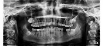
X-ray technology revolutionized medicine since its inception in 1895. Similar to the light we see every day, x-rays are a manifestation of electromagnetic radiation. Unlike light, however, they possess more photon energy–enough to penetrate and pass through material objects. In doing so, they can produce images of tissues and organs in the human (or non-human) body. If a detector is placed on the other side, it will capture those likenesses for medical examination. For much of radiology’s history, the film served as the primary detector. As with so many other areas, computers are assuming a much greater role as x-ray detectors.
How Do Digital X-Rays Differ from Traditional X-Rays?
Picture archive computer systems (also called PACS) capture x-rays and other images so that they can be transmitted and stored digitally in computer files that can be shared widely or closely. With traditional x-rays, the detector contains metallic film that must undergo development, as typical photographs once did. The process is both timely and costly. PACS detectors, by contrast, contain phosphor plates that are almost perpetually reusable (eventually paying for themselves).
Digital images, unlike those on film, are available in seconds. Furthermore, they do not need the physical storage space of film-based images which–another drawback–can degrade in quality over time. From an environmental standpoint, digital images do not need chemicals for development nor any byproduct to be disposed of. There are many advantages to PACS for doctors and dentists but patient safety certainly must be paramount in any diagnostic procedure.
Safety of Digital X-Rays
Because radiation is involved, many patients are wary of x-rays no matter the method and no matter the benefit. Scientific innovation speaks to this concern as the years unfold. Limiting the dosage of rays, employing lead shields and improving the film used each diminish the actual amount of radiation that enters the body. Yet digital radiography can cut down radiation by as much as 80 percent.
The reason behind this reduction is that digital phosphor detectors need much less exposure time, thereby limiting the quantity of radiation generated. Many dental patients undergo cleaning while x-rays are developed so they may not appreciate the speed and efficiency of digital radiography. Nevertheless, those concerned about the effect of radiation exposure can appreciate such a reduction in volume.
Health Plans Support Digital Radiography
Although not a perfect technology, digital radiography is endorsed by government insurance providers…in a back-handed way. Medicare reimbursements are now lower for x-ray procedures employing traditional means as opposed to digital equipment. As a result, digital radiography manufacturers are enjoying greater access to markets and higher stock prices. If this is any indication, there may be no going back to film.
Digital X-Rays
Laurel Dental Associates puts patient well-being front and center. Adopting healthy, safe, state-of-the-art technology like digital radiography is always based on diagnostic effectiveness with a minimum of side-effects. Given current results and future promise of digital x-rays, we will secure continued dental health…and your investment in it.


0 Comments
Comments are closed.